-
Die Universität
- Herzlich willkommen
- Das sind wir
- Medien & PR
-
Studium
- Allgemein
- Studienangebot
- Campusleben
-
Forschung
- Profil
- Infrastruktur
- Kooperationen
- Services
-
Karriere
- Arbeitgeberin Med Uni Graz
- Potenziale
- Arbeitsumfeld
- Offene Stellen
-
Diagnostik
- Patient*innen
- Zuweiser*innen
-
Gesundheitsthemen
- Gesundheitsinfrastruktur
Case of the Month
December 2022
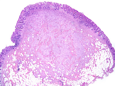
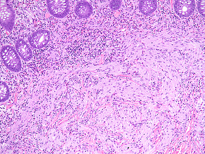
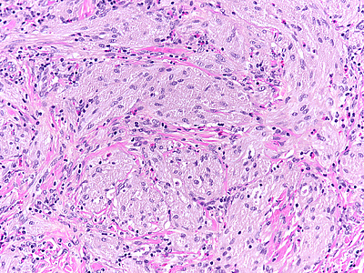
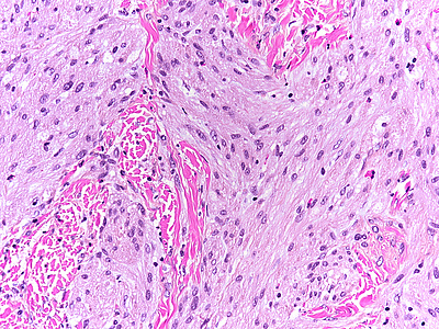
Colonic biopsy from a 55-year-old male.
Diagnosis
Granular cell tumour.
Comment
A 55-year-old male underwent endoscopy for large bowel cancer screening. A submucosal lesion was identified within the coecum and removed by snare polypectomy.
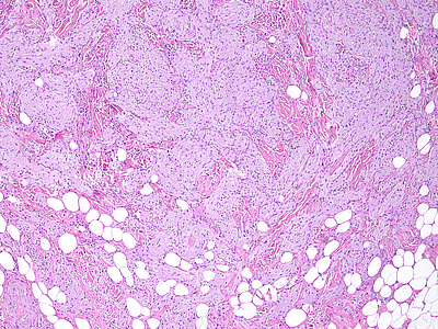
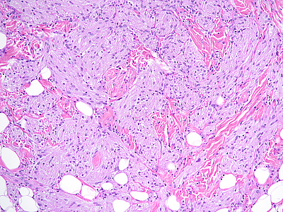
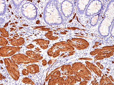
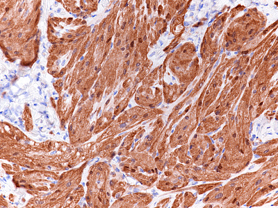
Histologically, we saw a submucosal proliferation of solid nests and ribbons of round to polyhedral cells, measuring approximately 6 mm in largest diameter (Panels A-B). The neoplastic cells generally contained small, uniform nuclei with inconspicuous nucleoli and abundant granular eosinophilic cytoplasm. They were separated by fibrous septa, which contained few inflammatory cells, mostly lymphocytes and few eosinophils (Panel C). In some areas, the neoplastic cells demonstrated cell spindling, still keeping the cytoplasmic features described above (Panel D). The lesion was ill-defined at the base, showing an infiltrative pattern into the surrounding adipose tissue within the submucosal layer (Panels E-F). The neoplastic cells were positive for PAS and strongly immunoreactive for S100-protein (Panels G-H), yet negative for keratin, CD117, DOG-1, and muscle markers, prompting final diagnosis of colonic granular cell tumour.
Granular cell tumors are common lesions in subcutaneous tissue. Gastrointestinal pathologists may encounter this form of peripheral nerve sheath tumour mainly in the oesophagus. In the colorectum, granular cell tumours prevail on the right side (coecum, around the ileocoecal valve, and within the ascending colon). On low power they are either infiltrative or well-defined, involving either the mucosa, submucosa (most common), or both. The cells are characteristically positive for PAS and S100-protein, but may also show positivity for SOX-10, NSE, and synaptophysin.
Behaviour is almost invariably benign, but patients may experience recurrence after incomplete excision. Reactive changes of the oerlying epithelium may be observed, mimicking a neoplastic process. This is an important pitfall within the oesophagus, but does not usually pose larger problems in the colorectum.
For further reading
- Singhi AD, Montgomery EA. Colorectal granular cell tumor: a clinicopathologic study of 26 cases. Am J Surg Pathol. 2010; 34: 1186-92. doi: 10.1097/PAS.0b013e3181e5af9d.
- Na JI, Kim HJ, Jung JJ, Kim Y, Kim SS, Lee JH, Lee KH, Park JT. Granular cell tumours of the colorectum: histopathological and immunohistochemical evaluation of 30 cases. Histopathology. 2014; 65: 764-74. doi: 10.1111/his.12487. Epub 2014 Aug 26.
- An S, Jang J, Min K, Kim MS, Park H, Park YS, Kim J, Lee JH, Song HJ, Kim KJ, Yu E, Hong SM. Granular cell tumor of the gastrointestinal tract: histologic and immunohistochemical analysis of 98 cases. Hum Pathol. 2015; 46: 813-9. doi: 10.1016/j.humpath.2015.02.005. Epub 2015 Feb 26.
- Chen Y, Chen Y, Chen X, Chen L, Liang W. Colonic granular cell tumor: Report of 11 cases and management with review of the literature. Oncol Lett. 2018; 16: 1419-1424. doi: 10.3892/ol.2018.8811. Epub 2018 May 25.
- Mobarki M, Dumollard JM, Dal Col P, Camy F, Peoc'h M, Karpathiou G. Granular cell tumor a study of 42 cases and systemic review of the literature. Pathol Res Pract. 2020; 216: 152865. doi: 10.1016/j.prp.2020.152865. Epub 2020 Feb 12.
Presented by
Dr. Cord Langner, Graz, and Dr. Stephan Bogner, Linz, Austria.


