-
Die Universität
- Herzlich willkommen
- Das sind wir
- Medien & PR
-
Studium
- Allgemein
- Studienangebot
- Campusleben
-
Forschung
- Profil
- Infrastruktur
- Kooperationen
- Services
-
Karriere
- Arbeitgeberin Med Uni Graz
- Potenziale
- Arbeitsumfeld
- Offene Stellen
-
Diagnostik
- Patient*innen
- Zuweiser*innen
-
Gesundheitsthemen
- Gesundheitsinfrastruktur
Case of the Month
March 2023
Three gastric polyps in a 59-year-old female.
Diagnosis
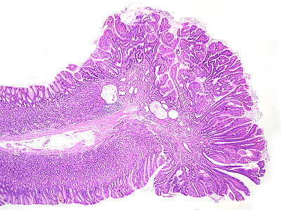
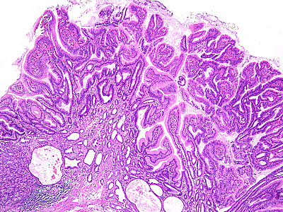
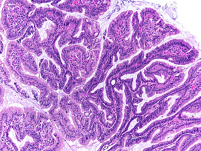
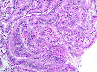
Sporadic foveolar-type gastric adenomas with a raspberry-like appearance.
Comment
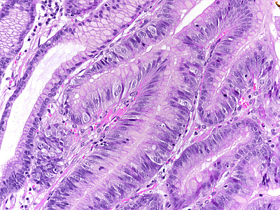
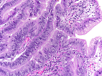
Three polyps, measuring from three to eight millimeters in maximum diameter, were incidentally discovered within the oxyntic mucosa of 59-year-old female and removed by cold snare polypectomy. There was no history of familial adenomatous polyposis (FAP), gastric adenocarcinoma and proximal polyposis of the stomach (GAPPS), or other inherited syndrome, as well as no personal or family history of cancer. No usage of PPI. The background mucosa was normal on gross inspection.
Upon histology, the three polyps showed a similar morphology. The polyps demonstrated irregularly shaped, branching, papillary projections with well-vascularized stroma (Panels A-C). On higher magnification, these projections were lined by dysplastic columnar cells, with hyperchromatic, enlarged and stratified nuclei and a distinctive apical mucin cap, prompting diagnosis of foveolar-type gastric adenoma with low-grade dysplasia (Panels D-F). The background tissue showed non-atrophic oxyntic mucosa, without gastritis, Helicobacter pylori infection or intestinal metaplasia. In summary, the histology was consistent with the newly described variant of foveolar-type adenoma with a raspberry-like appearance.
Broadly, gastric adenoma can be subdivided in four distinct entities: adenoma of intestinal type, adenoma of foveolar type, pyloric gland adenoma, and oxyntic gland adenoma. Of these, the foveolar-type gastric adenoma is histologically defined as a neoplastic epithelial polyp derived from gastric foveolar epithelium and showing foveolar cell differentiation, including positivity for MUC5AC. It develops in otherwise healthy gastric mucosa (no inflammation or atrophy/metaplasia) and is clinically classified into sporadic and syndromic types. The sporadic type is reported to be extremely rare, while the syndromic type occurs more frequently in patients with FAP or, rarely, GAPPS. Upon endoscopy, the foveolar-type gastric adenoma is usually described as slightly elevated whitish lesion, occurring in the oxyntic gastric compartment of the stomach.
Recently, a new histologic variant of foveolar-type gastric adenomas with a raspberry-like appearance has been reported. The study by Shibagaki et al. included 34 patients, seven of them with multiple lesions. The white-light endoscopy showed a small reddish polyp with a granular surface (raspberry-like appearance) and the narrow-band imaging with magnification endoscopy revealed an irregularly shaped papillary or gyrus-like superficial microstructure, with irregular or indistinct capillaries that were clearly demarcated from the surrounding mucosa, allowing the differential diagnosis from gastric hyperplastic polyp. The histological findings were as described in the case presented above.
Immunohistochemistry is not necessary for diagnosis. According to the publication, the neoplastic cells show strong and diffuse expression of MUC5AC, while they are negative or show only very low expression of MUC6, in particular at the basal portion of the neoplastic glands. Immunostaining for MUC2 and CD10 renders a negative result, thus exhibiting a pure gastric mucin phenotype (foveolar-cell differentiation). More studies are necessary, including molecular pathology, to elucidate the genetic profile of this peculiar new variant of foveolar-type adenoma, including potential differences between it and the traditional foveolar-type adenoma.
For further reading
- Shibagaki K, Mishiro T, Fukuyama C, Takahashi Y, Itawaki A, Nonomura S, Yamashita N, Kotani S, Mikami H, Izumi D, Kawashima K, Ishimura N, Nagase M, Araki A, Ishikawa N, Maruyama R, Kushima R, Ishihara S. Sporadic foveolar-type gastric adenoma with a raspberry-like appearance in Helicobacter pylori-naïve patients. Virchows Archiv (2021) 479:687-695. doi.org/10.1007/s00428-021-03124-3.
Presented by
Dr. Filipa Pereira, Lisbon, Portugal, and Dr. Cord Langner, Graz, Austria.


