-
Die Universität
- Herzlich willkommen
- Das sind wir
- Medien & PR
-
Studium
- Allgemein
- Studienangebot
- Campusleben
-
Forschung
- Profil
- Infrastruktur
- Kooperationen
- Services
-
Karriere
- Arbeitgeberin Med Uni Graz
- Potenziale
- Arbeitsumfeld
- Offene Stellen
-
Diagnostik
- Patient*innen
- Zuweiser*innen
-
Gesundheitsthemen
- Gesundheitsinfrastruktur
Case of the Month
May 2022
Small polyp in the sigmoid colon of a 75-year-old female.
Diagnosis
Schwann cell hamartoma.
Comment
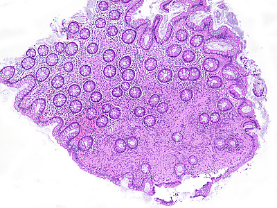
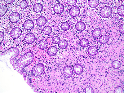
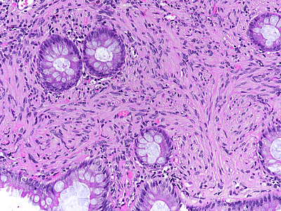
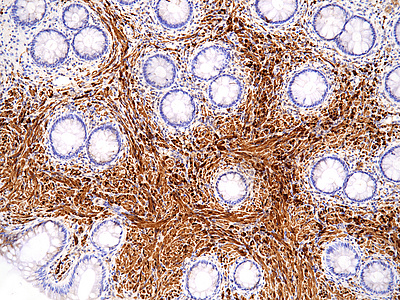
A dome-shaped sessile polypoid lesion with smooth glassy surface was removed from the sigmoid colon by forceps biopsy. Histology showed an expansion of the lamina propria by a diffuse proliferation of uniform bland-looking spindle cells with elongated tapering nuclei, abundant dense eosinophilic cytoplasm, and indistinct cell borders (Panels A-B). Colonic crypts were entrapped by the lesion. No nuclear atypia, pleomorphism, mitotic activity, or associated ganglion cells were observed (Panel C). The spindle cells were strong and diffusely positive for S-100 protein (Panel D).
Involvement of the gastrointestinal tract by benign nerve sheath tumours is rare. Neural lesions comprise a heterogeneous group of disorders, which may present as masses (such as schwannomas and neurofibromas) or, more commonly, as small polyps. The latter mainly include ganglioneuromas, perineuriomas, and granular cell tumours. Solitary diminutive polyps containing pure Schwann cell proliferations within the lamina propria have been identified as “mucosal Schwann cell hamartomas”. They mainly occur within the distal left colon and rectum, but may rarely also be detected in the upper gastrointestinal tract and gallbladder.
Differential diagnosis mainly includes NF1-associated neurofibromas, but may also include ganglioneuromas (ENGIP April 2017) and perineuriomas (ENGIP August 2014). The clinical significance of Schwann cell hamartomas is limited. Lesions are generally small and do not show progression to any other type of lesion.
For further reading
- Gibson JA, Hornick JL. Mucosal Schwann cell "hamartoma": clinicopathologic study of 26 neural colorectal polyps distinct from neurofibromas and mucosal neuromas. Am J Surg Pathol. 2009; 33: 781-787. doi: 10.1097/PAS.0b013e31818dd6ca.
- Pasquini P, Baiocchini A, Falasca L, Annibali D, Gimbo G, Pace F, Del Nonno F. Mucosal Schwann cell "Hamartoma": a new entity? World J Gastroenterol. 2009; 15: 2287-2289. doi: 10.3748/wjg.15.2287.
- Hytiroglou P, Petrakis G, Tsimoyiannis EC. Mucosal Schwann cell hamartoma can occur in the stomach and must be distinguished from other spindle cell lesions. Pathol Int. 2016; 66: 242-243. doi: 10.1111/pin.12376.
- Han J, Chong Y, Kim TJ, Lee EJ, Kang CS. Mucosal Schwann Cell Hamartoma in Colorectal Mucosa: A Rare Benign Lesion That Resembles Gastrointestinal Neuroma. J Pathol Transl Med. 2017; 51: 187-189. doi: 10.4132/jptm.2016.07.02.
- Chintanaboina J, Clarke K. Case of colonic mucosal Schwann cell hamartoma and review of literature on unusual colonic polyps. BMJ Case Rep. 2018 Sep 21;2018:bcr2018224931. doi: 10.1136/bcr-2018-224931.
- Vaamonde-Lorenzo M, Elorriaga K, Montalvo I, Bujanda L. Colonic mucosal Schwann cell hamartoma. J Dig Dis. 2020; 21: 475-477. doi: 10.1111/1751-2980.12874.
- Li Y, Beizai P, Russell JW, Westbrook L, Nowain A, Wang HL. Mucosal Schwann cell hamartoma of the gastroesophageal junction: A series of 6 cases and comparison with colorectal counterpart. Ann Diagn Pathol. 2020; 47: 151531. doi: 10.1016/j.anndiagpath.2020.151531.
Presented by
Dr. Ana Varelas, Porto, Portugal, and Dr. Cord Langner, Graz, Austria.


