-
Die Universität
- Herzlich willkommen
- Das sind wir
- Medien & PR
-
Studium
- Allgemein
- Studienangebot
- Campusleben
-
Forschung
- Profil
- Infrastruktur
- Kooperationen
- Services
-
Karriere
- Arbeitgeberin Med Uni Graz
- Potenziale
- Arbeitsumfeld
- Offene Stellen
-
Diagnostik
- Patient*innen
- Zuweiser*innen
-
Gesundheitsthemen
- Gesundheitsinfrastruktur
Case of the Month
June 2022
Biopsy material from the cardia in a 93-year-old male.
Diagnosis
Non-adenomatous (foveolar) dysplasia.
Comment
A 93-year-old male presented with unspecific abdominal discomfort. Upon upper endoscopy, the cardiac mucosa appeared polypoid and irregular. Multiple biopsies were taken.
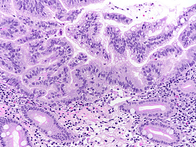
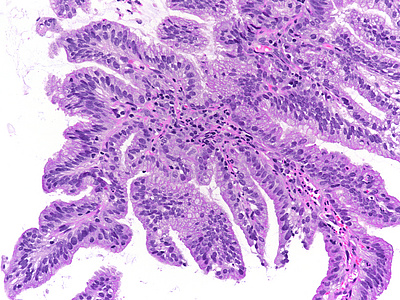
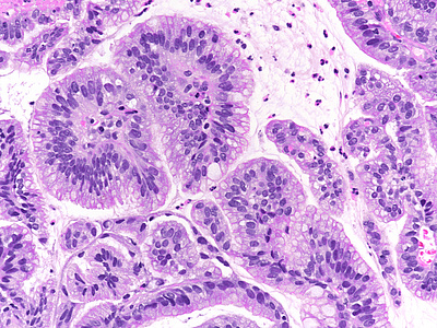
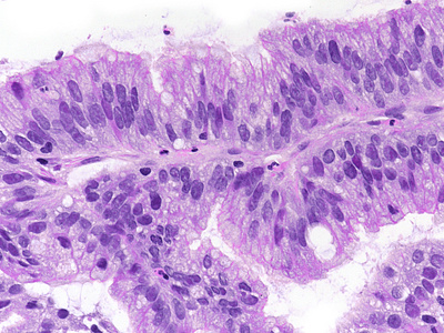
Histologically, the background mucosa revealed a chronic-atrophic gastritis with intestinal metaplasia (Panel A). The surface epithelium demonstrated a gastric foveolar phenotype with pale eosinophilic cytoplasm. The nuclei were oval, enlarged, but only slightly irregular. The chromatin content was increased and nuclear polarisation retained, respectively. Mitoses were inconspicuous (Panels A-D).
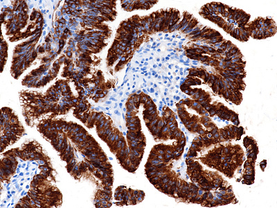
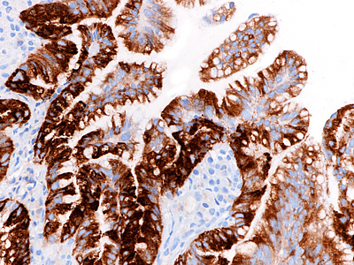
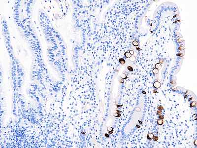
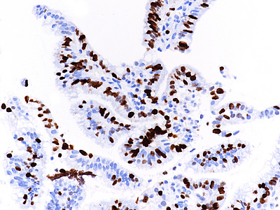
Immunohistochemistry confirmed the gastric foveolar phenotype, with positivity for MUC5AC (Panel E), MUC6 (Panel F) and negativity for MUC2 (Panel G). The Ki67 labelling index was 35% (Panel H).
The “intestinal” type of gastric carcinomas usually develops via an atrophy-metaplasia-dysplasia-carcinoma sequence. Traditionally, the gastric dysplasia occurring against a background of intestinal metaplasia shows an intestinal phenotype that recapitulates the morphology of colonic adenomas (MUC2 positive, MUC5AC and MUC6 negative). The same phenotype dominates the picture in Barrett’s dysplasia.
In recent years, several types of non-intestinal, that is, non-adenomatous dysplasia have been recognized. Of these, gastric foveolar dysplasia, as illustrated in our case, is the most frequent. It may be encountered within the stomach and at the gastro-esophageal junction, that is, in Barrett’s oesophagus. Gastric foveolar dysplasia is characterized by low columnar cells with clear / light eosinophilic cytoplasm (due to apical neutral mucin) and oval to round, often only slightly irregular nuclei.
It is of note that gastric foveolar dysplasia, even when appearing low grade, may give rise to very well differentiated adenocarcinomas. Therefore, the biopsy material needs to be examined thoroughly, e.g., by cutting multiple levels, not to overlook invasion. The inter-observer agreement in separating low-grade foveolar dysplasia from reactive gastric mucosa and low-grade adenomatous dysplasia appears to be poor. Greater awareness and agreed criteria are needed to prevent misdiagnosis of low-grade foveolar dysplasia as reactive, and vice versa.
Endoscopic removal of the dysplastic lesion by endoscopic mucosal resection (EMR) or endoscopic submucosal dissections (ESD) is the treatment of choice.
For further reading
- Khor TS, Alfaro EE, Ooi EM, Li Y, Srivastava A, Fujita H, Park Y, Kumarasinghe MP, Lauwers GY. Divergent expression of MUC5AC, MUC6, MUC2, CD10, and CDX-2 in dysplasia and intramucosal adenocarcinomas with intestinal and foveolar morphology: is this evidence of distinct gastric and intestinal pathways to carcinogenesis in Barrett Esophagus? Am J Surg Pathol. 2012; 36: 331-342. doi: 10.1097/PAS.0b013e31823d08d6.
- Patil DT, Bennett AE, Mahajan D, Bronner MP. Distinguishing Barrett gastric foveolar dysplasia from reactive cardiac mucosa in gastroesophageal reflux disease. Hum Pathol. 2013; 44: 1146-1153. doi: 10.1016/j.humpath.2012.10.004.
- Serra S, Chetty R. Non-adenomatous forms of gastro-oesophageal epithelial dysplasia: an under-recognised entity? J Clin Pathol. 2014; 67: 898-902. doi: 10.1136/jclinpath-2014-202600.
- Vieth M, Montgomery EA, Riddell RH. Observations of different patterns of dysplasia in barretts esophagus - a first step to harmonize grading. Cesk Patol. 2016; 52: 154-163.
- Serra S, Ali R, Bateman AC, Dasgupta K, Deshpande V, Driman DK, Gibbons D, Grin A, Hafezi-Bakhtiari S, Sheahan K, Srivastava A, Szentgyorgyi E, Vajpeyi R, Walsh S, Wang LM, Chetty R. Gastric foveolar dysplasia: a survey of reporting habits and diagnostic criteria. Pathology. 2017; 49: 391-396. doi: 10.1016/j.pathol.2017.01.007.
- Kővári B, Kim BH, Lauwers GY. The pathology of gastric and duodenal polyps: current concepts. Histopathology. 2021; 78: 106-124. doi: 10.1111/his.14275.
- Pereira D, Kővári B, Brown I, Chaves P, Choi WT, Clauditz T, Ghayouri M, Jiang K, Miller GC, Nakanishi Y, Kim KM, Kim BH, Kumarasinghe MP, Kushima R, Ushiku T, Yozu M, Srivastava A, Goldblum JR, Pai RK, Lauwers GY. Non-conventional dysplasias of the tubular gut: a review and illustration of their histomorphological spectrum. Histopathology. 2021; 78: 658-675. doi: 10.1111/his.14294.
- Kushima R. The updated WHO classification of digestive system tumours-gastric adenocarcinoma and dysplasia. Pathologe. 2022; 43: 8-15. doi: 10.1007/s00292-021-01023-7.
Presented by
Dr. Ana Varelas, Porto, Portugal, and Dr. Cord Langner, Graz, Austria.


