-
Die Universität
- Herzlich willkommen
- Das sind wir
- Medien & PR
-
Studium
- Allgemein
- Studienangebot
- Campusleben
-
Forschung
- Profil
- Infrastruktur
- Kooperationen
- Services
-
Karriere
- Arbeitgeberin Med Uni Graz
- Potenziale
- Arbeitsumfeld
- Offene Stellen
-
Diagnostik
- Patient*innen
- Zuweiser*innen
-
Gesundheitsthemen
- Gesundheitsinfrastruktur
Case of the Month
February 2024
Anal mass lesion in a 72-year-old female.
Diagnosis
Anal gland/duct-cyst.
Comment
A 72-year-old female presented with a cystic lesion of the anal canal, measuring 3 cm in largest diameter. Clinically, the lesion was suspected to represent an unusually large haemorrhoid node, and it was resected according to the Milligan-Morgan procedure.
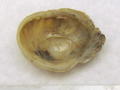
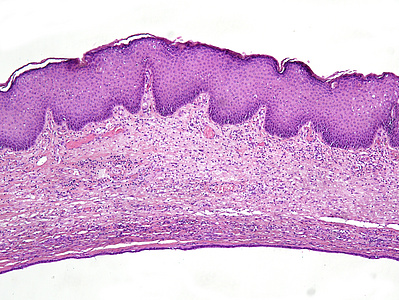
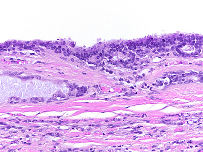
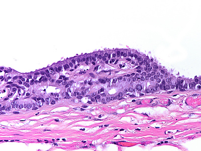
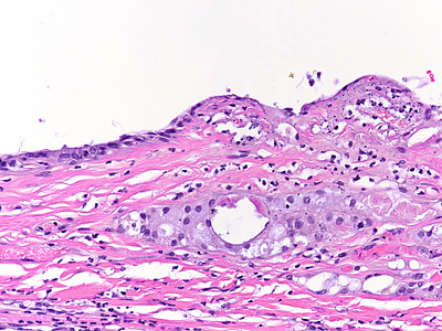
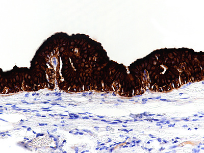
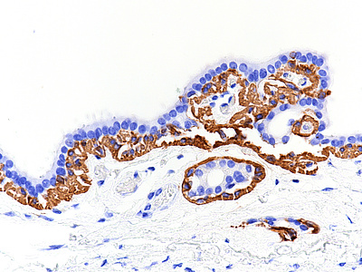
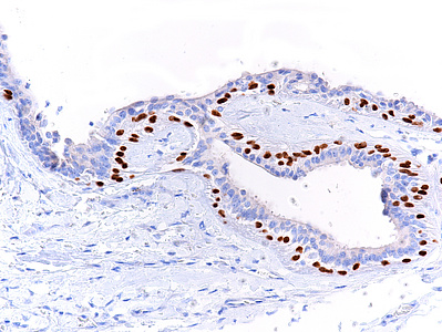
Upon gross inspection, we saw a cystic tumorous lesion with firm capsule and smooth inner surface, showing rare incomplete septa (Panel A). The cyst was lined by slightly disorganized, mainly flat cuboidal to columnar epithelium with abortive gland formation (Panels B-D). The nuclei were bland, with minor irregularities and small nucleoli in areas of active inflammation (Panel E). The lining epithelium was strongly positive for Keratin 7 (Panel F), albeit negative for Keratin 20 and the intestinal markers CDX-2 and SATB-2 (not shown). Keratin 5/6 (Panel G) and p40 (Panel H) disclosed a basal layer of cells with squamous features, which had been missed on H&E-stained slides.
Thus, the cyst recapitulated the morphology and immune profile of the anal glands and diagnosis of anal gland/duct-cyst was made. Though this entity is extremely rare, with only few case reports having been reported in the literature, the diagnosis is usually straight forward. Location within the transition zone of the anorectal region, with cyst lining epithelium displaying bidirectional, yet no colonic/rectal differentiation and no skin/adnexal differentiation, usually leads to the diagnosis.
Differential diagnosis of cystic lesions in the anrorectal region includes the tailgut cyst (retrorectal developmental cyst or cystic hamartoma), which has a different location and shows several types of epithelium (including ciliated, pure squamous and pure colonic/rectal epithelium), rectal mucocele (proctitis cystica profunda in mucosa prolapse syndrome) as well as cystic tumours of the perianal skin.
For further reading
- Kulaylat MN, Doerr RJ, Neuwirth M, Satchidanand SK. Anal duct/gland cyst: report of a case and review of the literature. Dis Colon Rectum. 1998; 41: 103-10.
- Seo GJ, Seo JH, Cho KJ, Cho HS. Anal Gland/Duct Cyst: A Case Report. Ann Coloproctol. 2020; 36: 204-206.
Presented by
Dr. Cord Langner, Graz, Austria.


