-
The University
- Welcome
- Who we are
- Media & PR
- Studying
-
Research
- Profile
- Infrastructure
- Cooperations
- Services
-
Career
- Med Uni Graz as an Employer
- Educational Opportunities
- Work Environment
- Job openings
-
Diagnostics
- Patients
- Referring physicians
-
Health Topics
- Health Infrastructure
Research team Brislinger
Research focus: Reproduction, pregnancy and regeneration
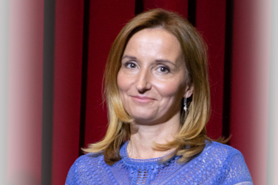
PI: Dagmar Brislinger
Focus: The importance of two-dimensional cell culture is non-controversial. Biochemical pathways and effects of treatments in single cell types can be identified in standardized processes. Nevertheless, the gap between a two-dimensional cell culture experiment and a complete organism is difficult to bridge, thus results can hardly be related. Although tissue and organ culture pose a challenge to the experimental design investigations will represent the in vivo situation closer than conventional two-dimensional cell culture. We therefore intensify and promote research on the establishment of three-dimensional cell cultures.
Network: The research group have established intense cooperation with researchers of the Gottfried Schatz Research Center for Cell Signaling, Metabolism and Aging and various institutes and divisions (e.g. Division of Transplantation Surgery, Division of Anaesthesiology for Cardiovascular Surgery and Intensive Care Medicine, Diagnostics & Research Institute of Pathology). Further, the research group has a long-lasting cooperation with the Institute of Multiphase Processes, Leibniz University Hannover, Germany.
Projects
Flexibility in Organ Research
- This project focuses on the development of various organ models – including a blood vessel model, a placenta model, an intestinal model, and a trachea model – using a 3D organ laboratory system. Existing three-dimensional systems or organ culture models often lack flexibility. In the model developed here, different cell types are embedded in a three-dimensional network composed of extracellular matrix. They can be cultured in defined compartments with distinct physical and chemical conditions, allowing direct cell-to-cell contact to be either promoted or prevented as needed..
- Duration: ongoing
- Funded by: Medical Universit Graz, FWF & CDG
- Project partner: Marc Müller, Institute of Multiphase Processes, Leibniz University Hannover, Germany
Isolation and characterization of endothelial cells and mesenchymal stem cells for vascular research and wound healing
- Mesenchymal stem/stromal cells (MSCs) are considered an ideal resource for clinical applications due to their low immunogenicity and relatively easy and abundant availability. We isolate, characterize, and culture MSCs from various human perinatal tissues (amnion, umbilical cord, chorionic vessels of the placenta) as well as from human adipose tissue. These MSCs are used either in single or co-culture with endothelial cells (isolated from the placenta or adult blood vessels) to seed vascular grafts, or are applied in a mouse model to study their effects on wound healing. Our previous in vitro and in vivo studies have shown that MSCs possess pro-angiogenic properties and enhance the viability and function of endothelial cells. Therefore, the application of MSCs in combination with biodegradable wound dressings represents a promising strategy to accelerate revascularization of wound areas.
- Duration: ongoing
- Funded by: Medical University Graz, COST (European Cooperation in Science and Technology)
- Project partner: Lars-Peter Kamolz, Klinische Abteilung für Plastische, Ästhetische und Rekonstruktive Chirurgie, Medizinische Universität Graz, Marc Müller, Institute of Multiphase Processes, Leibniz University Hannover, Germany
Innovative therapies in mucus associated diseases
- The mucus layers in the respiratory tract and the intestine form the major defense line between the host and the environment. Phytotherapy based on traditional Chinese medicine are increasingly used for various medical applications. In preliminary studies, we showed that a combination of selected Chinese herbal substances strongly affected the expression of gel-forming mucins in a cell culture model of the intestine and the respiratory tract. Thus, this project aims in the evaluation of herbal multi-compounds and the mechanistic basis for their regulatory elements in the mucus synthesis.
- Duration: ongoing
- Funded by: Med Uni Graz
- Project partners: Rudolf Bauer, Xuehong Nöst, Institute of Pharmaceutical Sciences, University of Graz, Austria
- PI: Dagmar Brislinger
Trophoblasts and glands in early human pregnancy
- We have demonstrated that fetal trophoblasts reach maternal uterine glands during implantation, that trophoblasts and glands interact early in pregnancy, and that trophoblasts invade the uterine glands (endoglandular trophoblast invasion), thus providing nutrition to the embryo before the establishment of uteroplacental circulation. Current projects examine endoglandular trophoblast invasion and the role of uterine glands and selected marker proteins from various perspectives, employing different techniques (histology, 3D culture models, organoids, bioinformatics, microbial effects, electron microscopy). A deeper understanding of endoglandular trophoblast invasion significantly expands current knowledge of early pregnancy. Using advanced in vitro models and targeted pharmacological/biomolecular modulation, we analyze the functional behavior of glandular epithelial cells and/or trophoblasts. Our goal is to further our understanding of the potential causes of fertility disorders and their treatment.
- Duration: ongoing
- Funded by: Medical University Graz, FWF, Acib-FFG-Comet
- Project partners: Sabine Kienesberger-Feist, Universität Graz, Bernhard Neumayer, FH Joanneum, eHealth
- PI: Gerit Moser
Building Better Blood Vessels: Human Protein Electrospun Scaffolds in Tissue Engineering
- The reconstruction of small-diameter blood vessels is a growing need due to the increasing prevalence of atherosclerotic diseases. While synthetic grafts perform well in large vessels, they are less suitable for smaller ones. Tissue-engineered, cell-seeded scaffolds offer a promising alternative. In this project, electrospun scaffolds made from human blood proteins are developed. The resulting nanofibrous 3D structure mimics the extracellular matrix (ECM), supporting strong cell adhesion and proliferation. The electrospinning technique enables continuous fiber production with diameters between 50 and 500 nm. To adapt this process to human plasma, previously established parameters using porcine blood must be modified. The patented method includes five steps: plasma separation, diafiltration, protein concentration, release of growth factors, and electrospinning. The scaffolds’ biocompatibility will be evaluated in vitro using endothelial cells and a chorioallantoic membrane (CAM) assay to assess angiogenic potential. The results of this project will provide a foundation for future research toward the development of tissue-engineered vascular grafts.
- Duration: 2024 - 2026
- Funded by: GESUNDHEIT 3000 Program of the MEFOgraz
- Project partner: Marc Müller, Institut für Mehrphasenprozesse, Leibniz Universität Hannover
- PI: Dagmar Brislinger
Uterine Trophoblast Invasion: A Matrix-Based 3D Vascular Model for Characterizing Pregnancy-Associated Diseases
- Successful embryo implantation and a healthy pregnancy depend on proper invasion of trophoblasts into maternal tissue. Disruptions in this process are linked to complications such as preeclampsia and intrauterine growth restriction. In early pregnancy, maternal arteries remain plugged by trophoblasts, while veins are already open and connected to the placenta — the reasons for this difference remain unclear. We hypothesize that endothelial factors play a key role. Due to the lack of suitable 3D models, we developed a novel, perfusable vessel model using a biodegradable PCL/PLA matrix, seeded with cells on both sides, creating two compartments with distinct culture conditions. The system enables co-culture of primary endothelial cells and first-trimester placental villi. Analyses combine traditional histological and biochemical techniques with advanced methods such as mRNA-based padlock probes and computer-assisted image analysis. Our goal is to gain new insights into trophoblast invasion and potential mechanisms underlying pregnancy-related disorders.
- Duration: 2025 - 2026
- Funded by: Unkonventionelle Forschung, Land Steiermark
- Project partners: Marc Müller, Institut für Mehrphasenprozesse, Leibniz Universität Hannover, Christian Baumgartner, Institut für Health Care Engineering mit Europaprüfstelle für Medizinprodukte, Technische Universität Graz
- PI MUG: Dagmar Brislinger
- PI TU: Julia Fuchs
Exogenous electrical and mechanical motion stimulation by using an electronic skin to support oral cell proliferation
- Periodontitis is a widespread disease affecting over 50% of adults in Europe, with severe forms causing tooth loss, impaired chewing, and reduced self-esteem. It is also linked to systemic conditions such as cardiovascular disease and diabetes, driven by chronic inflammation. This project explores the use of electronic skin (e-skin) for oral tissue regeneration. E-skin is a flexible, bioactive technology that mimics human skin and delivers targeted electrical and mechanical stimulation. By incorporating barium titanate (BaTiO₃) nanoparticles, we aim to enhance its mechanical properties, biocompatibility, and regenerative capacity. In phase 1, the bioactivity, cell adhesion, and stimulation potential of modified e-skin will be assessed. Phase 2 examines how electrical and mechanical cues affect gingival fibroblasts and rat bone marrow stromal cells (rBMSCs), which play key roles in regeneration via cytokine, chemokine, and exosome release. Phase 3 involves in vivo analysis of bone remodeling, a process influenced by mechanical and electrical stimuli. This controlled stimulation aims to significantly enhance tissue regeneration and offer new therapeutic strategies for treating periodontitis.
- Duration: 2025 - 2028
- Funded by: FWF Esprit
- Project partner: Uwe Yacine Schwarze, University Clinic for Dentistry and Oral Health and the Department of Orthopaedics and Traumatology, Medical University of Graz, Anna Maria Coclite, Department of Physics, University of Bari Aldo Moro, Italy
- PI: Fabiola Vaca de Marcher
Division of Cell Biology, Histology and Embryology
Team
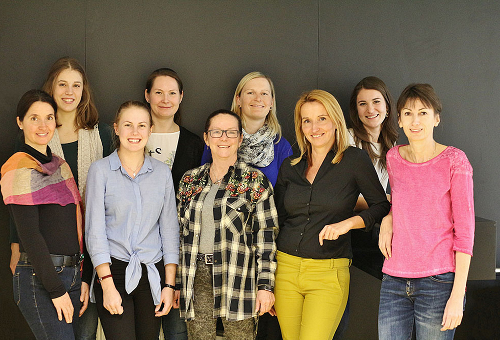
Members
- Fuchs Julia
- Jäger Lara
- Lang-Olip Ingrid
- Moser Gerit
- Pichlsberger Melanie
- Sundl Monika
- Kornhäusel Georg
- Moser Pia
- Scheiber Martina
- Vaca de Marcher Fabiola
- Wolf Janine


